
Chest X-ray and ECG
Learn from paired chest X-rays and 12-lead ECGs with the CardioVAE model [1], which can still make predictions when one of the two is missing.
Run this tutorial in Google Colab, which provides the GPU it uses for training.
In Colab, choose a T4 GPU runtime, then run Step 0 in order: install KaleCardiac and download the tutorial data. For Run locally, see Quick Start.
Sessions are temporary. Save code edits, important results and generated files before leaving or opening another example. Do not upload private or patient data to public services.
This tutorial uses KaleCardiac to pretrain, fine-tune, evaluate and interpret CardioVAE [1], a multimodal variational autoencoder (VAE) for two modalities: the chest X-ray (CXR) and the 12-lead electrocardiogram (ECG).
CardioVAE learns a shared latent representation of both modalities from paired, unlabelled ECGs and CXRs. Its tri-stream objective combines three evidence lower bounds (ELBOs): one through both modalities together and one through each modality alone, so that each encoder remains useful when the other modality is missing. The modalities are fused at the feature-embedding level (intermediate fusion), and a classifier is then fine-tuned on top of the pretrained encoders.
The workflow starts from raw DICOM files, the format in which hospitals export ECGs and chest X-rays, and each step takes a few calls to the KaleCardiac API:
Step 0: Environment preparation. Install KaleCardiac and download the tutorial dataset.
Step 1: Multimodal pretraining. Load the pretraining configuration, load and prepare the unlabelled ECG–CXR pairs, then pretrain CardioVAE.
Step 2: Multimodal fine-tuning. Load the fine-tuning configuration, load and prepare the labelled pairs, then fine-tune a classifier.
Step 3: Evaluation. Evaluate the held-out test subjects.
Step 4: Interpretation. Identify which modality, and which regions of each ECG and CXR, the predictions rely on.
Estimated runtime: about 30 minutes on a Colab T4 GPU, including installation. The estimates for individual steps below also refer to a T4 GPU.
Step 0: Environment Preparation
Runtime
In Google Colab, select Runtime > Change runtime type > T4 GPU. The tutorial also runs on a CPU, more slowly.
Package installation
The cell below installs KaleCardiac from PyPI, with two optional extras: imaging (pydicom and scikit-image, required by PyKale’s DICOM reader) and interpret (Captum, for integrated gradients). Each configuration file is downloaded later, by the cell that reads it.
Run this cell in Google Colab. When running locally, skip it if KaleCardiac is already installed, for example with pip install -e ".[imaging,interpret]" from a clone of its repository; opened from that clone’s tutorials/mdas_tutorial folder, the notebook reads the configuration files there.
Estimated runtime: 3 minutes
!pip install --quiet "kalecardiac[imaging,interpret]"
Setup
The cell below silences warnings and PyTorch Lightning’s start-up messages to keep the output readable, then reports the installed KaleCardiac version and the working directory, confirming that the installation succeeded.
import logging
import warnings
from pathlib import Path

import pytorch_lightning

import kalecardiac

warnings.filterwarnings("ignore")
# Importing Lightning sets its loggers' level, so they are quieted after it is imported.
for name in (pytorch_lightning.__name__, "lightning_fabric"):
    logging.getLogger(name).setLevel(logging.ERROR)
print(f"KaleCardiac {kalecardiac.__version__}, working in {Path.cwd()}")
KaleCardiac 0.1.0a2, working in M:\content
Dataset download
The tutorial dataset is a single ZIP archive. The cell below downloads it into the runtime’s local storage, without any sign-in, and unpacks it into data/ next to this notebook:
data/
├── kalecardiac_pretraining/
│   ├── ECG/<subject>/.../*.dcm    one twelve-lead ECG per subject (DICOM ECG waveform)
│   └── CXR/<subject>/.../*.dcm    one chest X-ray per subject (DICOM radiograph)
└── kalecardiac_finetuning/
    ├── ECG/<subject>/.../*.dcm
    ├── CXR/<subject>/.../*.dcm
    └── labels.csv                 subject_id,label
kalecardiac_pretraining holds unlabelled ECG–CXR pairs for pretraining; kalecardiac_finetuning adds a label for each subject for fine-tuning. As in a hospital export, each subject has a folder named by its identifier, and a subject’s ECG and CXR are paired by that name. To use your own data instead, skip this cell and see Your own data below.
Estimated runtime: 2 minutes
import urllib.request
import zipfile

# The tutorial dataset on Dropbox; "dl=1" asks Dropbox for the file itself rather than its web page.
DATA_URL = "https://www.dropbox.com/scl/fi/y488iouenrk641dxtd8ai/kalecardiac_tutorial_data.zip?rlkey=rorf5mvbt24jdw2cr3ecyy91n&st=yt83r37g&dl=1"

if not Path("data").is_dir():
    archive, _ = urllib.request.urlretrieve(DATA_URL, "kalecardiac_tutorial_data.zip")
    with zipfile.ZipFile(archive) as bundle:
        bundle.extractall("data")
print(sorted(folder.name for folder in Path("data").iterdir()))
['kalecardiac_finetuning', 'kalecardiac_pretraining']
Your own data
The same steps run on your own ECGs and chest X-rays, organised as in the tutorial dataset: ECG/ and CXR/ folders, each with one folder per subject containing that subject’s DICOM file, and, for fine-tuning, a labels.csv file with a subject_id column and a column of classes (0 or 1). Skip the dataset download, and set your folders in the configuration cells of Step 1 and Step 2 by uncommenting the lines marked Your own data. Sampling rates are read from the DICOM headers, and recordings are resampled to the 500 Hz that CardioVAE reads.
Use identifiable patient data only in an approved, secure environment, never on public services such as Colab.
Step 1: Multimodal Pretraining
CardioVAE is pretrained without labels on the paired ECGs and CXRs in kalecardiac_pretraining.
Configuration
KaleCardiac reads every setting from a YACS configuration, which keeps an experiment’s settings separate from its code. The tutorial’s configuration files are in the configs folder of its folder in the KaleCardiac repository, and each is merged onto KaleCardiac’s defaults (kalecardiac.config). The cell below downloads the pretraining file, config_cardiovae_pretrain.yaml, into a configs folder beside the notebook, unless one is already there, and reads it: the unlabelled dataset, the ECG and CXR input shapes (including the order of the ECG leads), the latent width, the tri-stream objective and the pretraining schedule.
Change a setting in the file, or override it in the cell below.
import urllib.request

from kalecardiac.config import get_cfg_defaults

# The tutorial's configuration files, as released with the KaleCardiac version installed above.
CONFIG_URL = f"https://raw.githubusercontent.com/pykale/cardiac/v{kalecardiac.__version__}/tutorials/mdas_tutorial/configs"
config_pt = Path("configs/config_cardiovae_pretrain.yaml")
if not config_pt.is_file():  # a copy already here, such as one you have edited, is kept
    config_pt.parent.mkdir(exist_ok=True)
    urllib.request.urlretrieve(f"{CONFIG_URL}/{config_pt.name}", config_pt)

cfg_pt = get_cfg_defaults()
cfg_pt.merge_from_file(config_pt)

# ------ Hyperparameters to play with -----
cfg_pt.MODEL.LATENT_DIM = 128
cfg_pt.SOLVER.MAX_EPOCHS = 100
cfg_pt.SOLVER.BATCH_SIZE = 4
cfg_pt.SOLVER.BASE_LR = 1e-4
cfg_pt.OBJECTIVE.RECONSTRUCTION_WEIGHTS = ["ecg:10", "cxr:1"]
cfg_pt.OBJECTIVE.ANNEALING_EPOCHS = cfg_pt.SOLVER.MAX_EPOCHS  # the KL weight rises over the whole run

# ------ Your own data -----
# cfg_pt.DATASET.ROOT = "/path/to/your/pretraining_data"  # holds ECG/ and CXR/

print(f"DATASET.ROOT: {cfg_pt.DATASET.ROOT}\n")
for section in ("ECG", "IMAGE", "OBJECTIVE", "SOLVER"):
    print(f"{section}:\n{cfg_pt[section]}\n")
DATASET.ROOT: data/kalecardiac_pretraining

ECG:
ENCODER: conv
LEADS: ['I', 'II', 'III', 'aVR', 'aVF', 'aVL', 'V1', 'V2', 'V3', 'V4', 'V5', 'V6']
LENGTH: 60000
NORMALIZE: True
NUM_CHANNELS: 1
SAMPLING_RATE: 500
SOURCE_SAMPLING_RATE: 0

IMAGE:
CHANNELS: 1
SCALE: True
SIZE: [224, 224]

OBJECTIVE:
ALIGNMENT_WEIGHT: 0.0
ANNEALING_EPOCHS: 100
POS_WEIGHT: 0.0
RECONSTRUCTION_WEIGHTS: ['ecg:10', 'cxr:1']
SCALE_FACTOR: 0.0001
UNIMODAL_STREAMS: True

SOLVER:
ACCELERATOR: auto
BASE_LR: 0.0001
BATCH_SIZE: 4
DEVICES: auto
EARLY_STOP: 0
GRAD_CLIP: 1.0
MAX_EPOCHS: 100
OPTIMIZER: Adam
SEED: 123
WEIGHT_DECAY: 0.0
Data loading
ECGFileSource.from_dicom and ImageFileSource.from_dicom, in kalecardiac.loaddata, index each modality’s folder by subject and read a subject’s file only when it is needed, so cohorts of any size load in the same way. Each file is read with PyKale’s [3] DICOM reader, kale.loaddata.image_access.read_dicom_phases, and then:
ECG: the 12-lead rhythm waveform is extracted in millivolts, with each lead’s name and the sampling rate read from the file’s header. Leads are matched by name, so a file that stores aVF before aVL cannot transpose them.
CXR: the pixel values are mapped through the display settings in the file’s header (the rescale, the window and, for
MONOCHROME1images, the inversion), so the model sees the image as a clinician does.
Estimated runtime: 10 seconds
from kalecardiac.loaddata import ECGFileSource, ImageFileSource

ecg_dir_pt, cxr_dir_pt = Path(cfg_pt.DATASET.ROOT, "ECG"), Path(cfg_pt.DATASET.ROOT, "CXR")
ecg_raw = ECGFileSource.from_dicom(ecg_dir_pt)
cxr_raw = ImageFileSource.from_dicom(cxr_dir_pt)
print(ecg_raw, cxr_raw, sep="\n")
ECGFileSource(20 subjects | leads=['I', 'II', 'III', 'aVR', 'aVL', 'aVF', 'V1', 'V2', 'V3', 'V4', 'V5', 'V6'] | shape=12x5120 | 500 Hz)
ImageFileSource(20 subjects | 1x2634x2643)
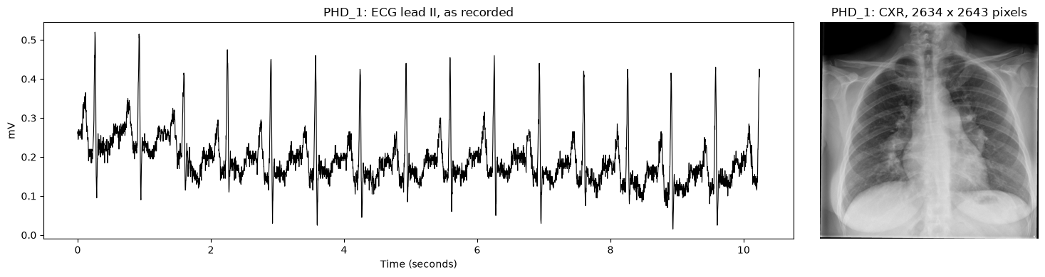
One subject’s ECG and CXR as loaded: lead II of the full recording, and the radiograph at its original resolution.
import matplotlib.pyplot as plt
import numpy as np

subject = sorted(ecg_raw.paths)[0]
recording, radiograph = ecg_raw.get(subject, 0).numpy(), cxr_raw.get(subject, 0).numpy()[0]

fig, (ax_ecg, ax_cxr) = plt.subplots(1, 2, figsize=(15, 4), gridspec_kw={"width_ratios": [3, 1]})
time = np.arange(recording.shape[1]) / ecg_raw.sampling_rate
ax_ecg.plot(time, recording[ecg_raw.leads.index("II")], color="black", linewidth=0.8)
ax_ecg.set(xlabel="Time (seconds)", ylabel="mV", title=f"{subject}: ECG lead II, as recorded")
ax_cxr.imshow(radiograph, cmap="gray")
ax_cxr.set_title(f"{subject}: CXR, {radiograph.shape[0]} x {radiograph.shape[1]} pixels")
ax_cxr.axis("off")
plt.tight_layout()
plt.show()
Data preparation
For each subject, CardioVAE reads ten seconds of the 12-lead ECG recorded at 500 Hz, which gives 5,000 time points per lead, and a 224 × 224 CXR scaled to [0, 1]. The 12-leads are interleaved time point by time point into a single strip of 60,000 values (12 leads × 5,000 time points). Two KaleCardiac transforms build these inputs from the configuration:
get_ecg_transform_from_cfg(kalecardiac.auto) readscfg.ECG: it fills any missing values in the signal, resamples a recording made at another rate to 500 Hz, crops or pads each lead to ten seconds, normalizes each lead to zero mean and unit variance, and then interleaves the leads. Every step before the interleaving works on one lead at a time. The leads are taken by name, in the order ofECG.LEADS: the order in which the published CardioVAE was pretrained, with aVF before aVL.get_image_transform(kalecardiac.prepdata) resizes the CXR with anti-aliasing and scales it to [0, 1].
The sources apply these transforms as each subject is read. MultimodalDataset then pairs the two modalities by subject: each subject yields its ECG as ecg and its CXR as cxr, the names used by the objective’s weights and the model’s encoders.
Estimated runtime: 10 seconds
from kalecardiac.auto import get_ecg_transform_from_cfg
from kalecardiac.loaddata import MultimodalDataset
from kalecardiac.prepdata import get_image_transform

# The rate the recordings were made at, as their DICOM headers state it, so any other is resampled.
cfg_pt.ECG.SOURCE_SAMPLING_RATE = ecg_raw.sampling_rate
ecg_transform = get_ecg_transform_from_cfg(cfg_pt)
cxr_transform = get_image_transform(size=cfg_pt.IMAGE.SIZE)

ecg = ECGFileSource.from_dicom(ecg_dir_pt, select=cfg_pt.ECG.LEADS, transform=ecg_transform)
cxr = ImageFileSource.from_dicom(cxr_dir_pt, transform=cxr_transform)
pretrain_set = MultimodalDataset(sorted(ecg.paths.keys() & cxr.paths.keys()), {"ecg": ecg, "cxr": cxr})

print(pretrain_set)
print({name: tuple(value.shape) for name, value in pretrain_set[0].modalities.items()})
MultimodalDataset(20 subjects | ecg=ECGFileSource, cxr=ImageFileSource)
{'ecg': (1, 60000), 'cxr': (1, 224, 224)}
Multimodal pretraining
CardioVAE, in kalecardiac.model.embed, has an encoder and a decoder for each modality: a 1-D convolutional network for the ECG strip and a 2-D convolutional network for the CXR. It fuses the modalities with a product of experts: each encoder’s Gaussian over the shared latent space and a standard-normal prior are multiplied into one joint posterior, and a subject missing a modality contributes no expert for it. The input sizes are taken from the prepared data.
pretrain, in kalecardiac.auto, trains the model with the configured objective (the tri-stream ELBO, with the ECG’s reconstruction weighted ten times the CXR’s and the KL term annealed in) and saves the weights to pretrained.pt in OUTPUT.OUT_DIR. Pretraining is a reconstruction task, so it needs no labels.
Estimated runtime: 10 to 20 minutes
import pandas as pd

from kalecardiac.auto import pretrain
from kalecardiac.model.embed import CardioVAE
from kalecardiac.utils import set_seed

set_seed(cfg_pt.SOLVER.SEED)
model = CardioVAE(
    signal_length=ecg.length,
    image_size=cxr.size,
    latent_dim=cfg_pt.MODEL.LATENT_DIM,
    signal_name="ecg",
    image_name="cxr",
)
print(f"CardioVAE with {sum(parameter.numel() for parameter in model.parameters()):,} parameters")

pretrained = pretrain(cfg_pt, model, pretrain_set, cfg_pt.OUTPUT.OUT_DIR)

# The training loss per epoch, from the run's log.
log = max(Path(cfg_pt.OUTPUT.OUT_DIR).glob("pretrain/history/*/metrics.csv"), key=lambda path: path.stat().st_mtime)
pd.read_csv(log).groupby("epoch")["train_loss"].mean().round(1).to_frame("training loss").T
CardioVAE with 204,180,418 parameters
| epoch | 0 | 1 | 2 | 3 | 4 | 5 | 6 | 7 | 8 | 9 | ... | 90 | 91 | 92 | 93 | 94 | 95 | 96 | 97 | 98 | 99 |
|---|---|---|---|---|---|---|---|---|---|---|---|---|---|---|---|---|---|---|---|---|---|
| training loss | 189.9 | 189.1 | 189.1 | 188.8 | 188.2 | 187.8 | 187.4 | 186.9 | 186.5 | 186.1 | ... | 176.8 | 176.9 | 176.7 | 176.4 | 176.7 | 176.7 | 176.7 | 176.5 | 176.2 | 176.3 |
1 rows × 100 columns
Step 2: Multimodal Fine-tuning
The pretrained encoders are fine-tuned on the labelled pairs in kalecardiac_finetuning to classify each subject: 0 healthy, 1 cardiothoracic abnormality.
Configuration
Fine-tuning starts from the pretrained model, so its configuration file, config_cardiovae_finetune.yaml, which the cell below downloads as the pretraining one was, is merged onto the pretraining configuration. It adds the labelled dataset, the endpoint, the train/validation/test split and the fine-tuning schedule.
Change a setting in the file, or override it in the cell below.
# The fine-tuning configuration file, downloaded as the pretraining one was.
config_ft = Path("configs/config_cardiovae_finetune.yaml")
if not config_ft.is_file():
    urllib.request.urlretrieve(f"{CONFIG_URL}/{config_ft.name}", config_ft)

# Fine-tuning starts from the pretrained model, and so from its configuration.
cfg_ft = cfg_pt.clone()
cfg_ft.merge_from_file(config_ft)

# ------ Hyperparameters to play with -----
cfg_ft.FINETUNE.MAX_EPOCHS = 100
cfg_ft.FINETUNE.BATCH_SIZE = 4
cfg_ft.MODEL.HEAD_HIDDEN = [128]

# ------ Your own data -----
# cfg_ft.DATASET.ROOT = "/path/to/your/finetuning_data"  # holds ECG/, CXR/ and labels.csv
# cfg_ft.ENDPOINT.LABEL_COLUMN = "label"  # the column of labels.csv holding each subject's class

print(f"DATASET.ROOT: {cfg_ft.DATASET.ROOT}")
print(f"ENDPOINT: {cfg_ft.ENDPOINT.NAME}, the {cfg_ft.ENDPOINT.LABEL_COLUMN!r} column of labels.csv\n")
print(f"FINETUNE:\n{cfg_ft.FINETUNE}")
DATASET.ROOT: data/kalecardiac_finetuning
ENDPOINT: cardiothoracic_abnormality, the 'label' column of labels.csv

FINETUNE:
BASE_LR: 0.001
BATCH_SIZE: 4
MAX_EPOCHS: 100
Data loading
The labelled cohort has the pretraining layout plus labels.csv, with one row per subject. Its ECGs and CXRs load through the same DICOM sources and transforms, because the pretrained encoders expect exactly these input shapes; the labels are an ordinary table.
Estimated runtime: 1 second
ecg_ft = ECGFileSource.from_dicom(Path(cfg_ft.DATASET.ROOT, "ECG"), select=cfg_pt.ECG.LEADS, transform=ecg_transform)
cxr_ft = ImageFileSource.from_dicom(Path(cfg_ft.DATASET.ROOT, "CXR"), transform=cxr_transform)
labels = pd.read_csv(Path(cfg_ft.DATASET.ROOT, "labels.csv"))

print(ecg_ft, cxr_ft, sep="\n")
labels.head()
ECGFileSource(20 subjects | leads=['I', 'II', 'III', 'aVR', 'aVF', 'aVL', 'V1', 'V2', 'V3', 'V4', 'V5', 'V6'] | shape=1x60000 | 500 Hz)
ImageFileSource(20 subjects | 1x224x224)
| subject_id | label | |
|---|---|---|
| 0 | PHD_1 | 0 |
| 1 | PHD_2 | 1 |
| 2 | PHD_3 | 0 |
| 3 | PHD_4 | 1 |
| 4 | PHD_5 | 0 |
Data preparation
Three calls, each reading cfg_ft, turn the labels into a fine-tuning task:
derive_endpointreads each subject’s class fromENDPOINT.LABEL_COLUMN, dropping a subject without a label rather than assuming it is negative.get_endpoint_targetmakes the class the supervision target that every batch carries.get_foldsdivides the subjects into training, validation and test sets, stratified by label, with no subject in two sets. 20% of the subjects are held out for testing, and the validation set that selects the checkpoint is drawn from the rest, never from the test set.
The class weight used by the loss is computed from the training set alone.
Estimated runtime: 1 second
from kalecardiac.auto import derive_endpoint, get_endpoint_target, get_folds

table = derive_endpoint(cfg_ft, labels)
target = get_endpoint_target(cfg_ft, table, id_column="subject_id")
fold = get_folds(cfg_ft, table)[0]
print(
    f"{len(fold.train_ids)} subjects to train on, {len(fold.val_ids)} to validate on, {len(fold.test_ids)} to test on"
)
13 subjects to train on, 3 to validate on, 4 to test on
Multimodal fine-tuning
fine_tune, in kalecardiac.auto, keeps the pretrained CardioVAE encoders frozen as feature extractors and trains a classification head on the concatenated posterior means of both modalities. The head, the loss and the positive class’s weight follow from the endpoint and the training labels. After training, the checkpoint with the best validation score is restored and the test subjects are scored.
Estimated runtime: 5 to 10 minutes
from kalecardiac.auto import fine_tune

set_seed(cfg_ft.SOLVER.SEED)
result = fine_tune(cfg_ft, pretrained, {"ecg": ecg_ft, "cxr": cxr_ft}, target, fold, cfg_ft.OUTPUT.OUT_DIR)
Step 3: Evaluation
fine_tune evaluates the held-out test subjects with score_predictions, which chooses the metrics suited to the endpoint; for a binary endpoint, these come from binary_metrics in kalecardiac.evaluate:
Accuracy: the proportion of subjects classified correctly.
AUROC: the area under the receiver operating characteristic curve, which measures how well the scores rank positive subjects above negative ones across all thresholds.
MCC: the Matthews correlation coefficient, which remains informative when the classes are imbalanced.
Sensitivity and specificity: the proportions of positive and of negative subjects classified correctly.
AUROC does not depend on a decision threshold, whereas accuracy, MCC, sensitivity and specificity are computed at a threshold of 0.5, so a model can rank subjects well while its scores still fall on one side of that threshold. The predictions are kept for each subject, so they can be inspected or scored again at another threshold.
Note: The tutorial dataset is synthetic and very small, so these scores only show that the API runs, not how the model would perform on real data.
metrics = pd.Series(result.metrics)[["accuracy", "roc_auc", "mcc", "sensitivity", "specificity"]].astype(float)
metrics.rename({"roc_auc": "AUROC", "mcc": "MCC"}).round(3).to_frame("test")
| test | |
|---|---|
| accuracy | 0.750 |
| AUROC | 1.000 |
| MCC | 0.577 |
| sensitivity | 1.000 |
| specificity | 0.500 |
predictions = pd.DataFrame(result.predictions.rows())
predictions.assign(probability=1 / (1 + np.exp(-predictions["score"]))).round(3)
| subject_id | split | fold | score | label | probability | |
|---|---|---|---|---|---|---|
| 0 | PHD_13 | test | 0 | 0.098 | 0.0 | 0.525 |
| 1 | PHD_15 | test | 0 | 1.385 | 1.0 | 0.800 |
| 2 | PHD_6 | test | 0 | 1.385 | 1.0 | 0.800 |
| 3 | PHD_7 | test | 0 | -2.020 | 0.0 | 0.117 |
Step 4: Interpretation
Two questions, both answered from the fine-tuned model:
Which modality does the model rely on?
interpret_model, inkalecardiac.auto, runs integrated gradients over the test subjects and reports each modality’s attribution ratio: the share of its ECG time points or CXR pixels that are strongly attributed. It then withholds each modality in turn, through the presence mask, and scores the test set again; a modality whose removal costs little is one the model can do without.Where in each modality?
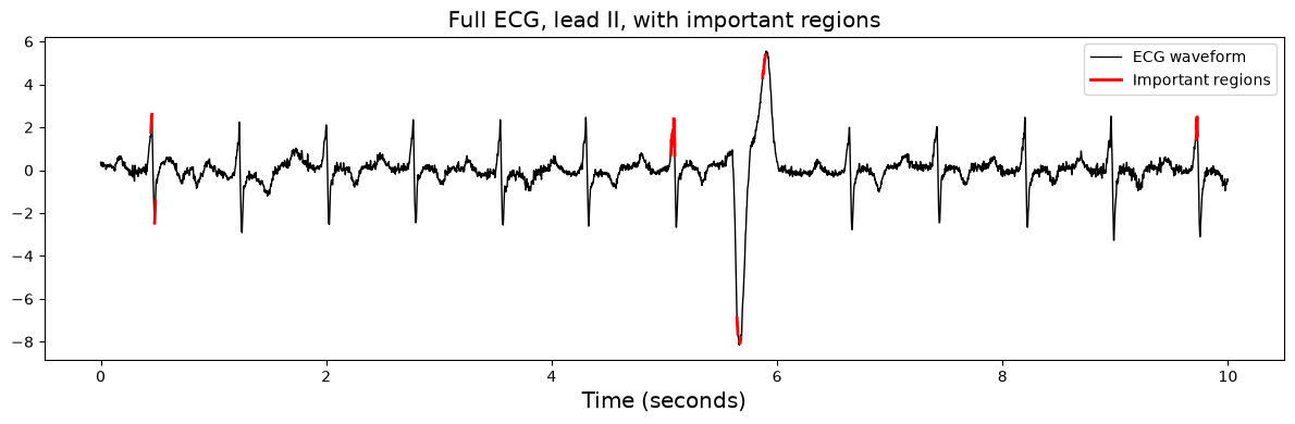
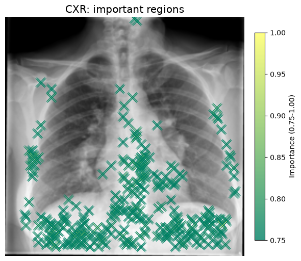
modality_attributions, inkalecardiac.interpret, attributes one subject’s prediction to every time point of its ECG and every pixel of its CXR with Captum’s integrated gradients [2], andnormalize_attributionrescales each map to [0, 1], so that the regions above a threshold can be highlighted.
Estimated runtime: 10 seconds
from kalecardiac.auto import interpret_model

summary = interpret_model(cfg_ft, result.model, result.test_loader, cfg_ft.OUTPUT.OUT_DIR)
pd.DataFrame(summary["attribution"]).T.round(3)
| ratio | share | |
|---|---|---|
| ecg | 0.014 | 0.556 |
| cxr | 0.011 | 0.444 |
The test AUROC with every modality (full), and how far it falls when each modality is withheld (drop_ecg, drop_cxr):
# A drop is the full score minus the score without that modality.
ablation = summary.get("ablation", {})
drops = {name: score for name, score in ablation.items() if not name.startswith("without_")}
pd.Series(drops, name="test AUROC").round(3).to_frame()
| test AUROC | |
|---|---|
| full | 1.0 |
| drop_ecg | 0.5 |
| drop_cxr | 0.0 |
One test subject’s prediction, attributed to its ECG and CXR. The ECG strip interleaves the twelve leads, so a single lead is read out of it by name.
import torch

from kalecardiac.interpret import modality_attributions, normalize_attribution
from kalecardiac.loaddata import collate_subjects

# Change these to explain another test subject or lead, or to move the thresholds.
SAMPLE_IDX, LEAD = 0, "II"
ECG_THRESHOLD, CXR_THRESHOLD, ZOOM_RANGE = 0.75, 0.75, (3.5, 4.0)

sample = collate_subjects([result.test_loader.dataset[SAMPLE_IDX]])
attributions = modality_attributions(result.model.model, sample.modalities, present=sample.present)

# The strip interleaves the leads time point by time point: one row per time point, one column per lead.
lead, num_leads = ecg_ft.leads.index(LEAD), len(ecg_ft.leads)
ecg_signal = sample.modalities["ecg"][0, 0].numpy().reshape(-1, num_leads)[:, lead]
ecg_importance = normalize_attribution(attributions["ecg"][0, 0]).reshape(-1, num_leads)[:, lead]
ecg_time = np.arange(ecg_signal.size) / cfg_ft.ECG.SAMPLING_RATE
cxr_image = sample.modalities["cxr"][0, 0].numpy()
cxr_importance = normalize_attribution(attributions["cxr"][0, 0])

probability = torch.sigmoid(result.model.predict(sample).prediction).item()
print(
    f"{sample.subject_id[0]}: label {int(sample.target['label'])}, predicted probability of a cardiothoracic "
    f"abnormality {probability:.2f}"
)
PHD_13: label 0, predicted probability of a cardiothoracic abnormality 0.52
Full ECG view: the full ten seconds of one lead, with the time points whose attribution exceeds the threshold drawn in red.
def plot_ecg(window: slice, title: str, width: float = 1.0):
    fig, ax = plt.subplots(figsize=(12, 4))
    ax.plot(ecg_time[window], ecg_signal[window], color="black", linewidth=width)
    for index in np.flatnonzero(ecg_importance[window] >= ECG_THRESHOLD):
        stretch = slice(max(index - 2, 0), index + 3)
        ax.plot(ecg_time[window][stretch], ecg_signal[window][stretch], color="red", linewidth=2 * width)
    ax.set_xlabel("Time (seconds)", fontsize="x-large")
    ax.set_title(title, fontsize="x-large")
    ax.legend(["ECG waveform", "Important regions"], fontsize="medium")
    plt.tight_layout()
    plt.show()


plot_ecg(slice(None), f"Full ECG, lead {LEAD}, with important regions")
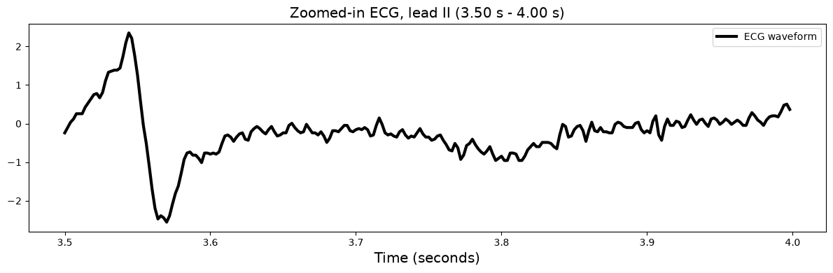
Zoomed-in ECG segment: a short stretch of the same lead, 3.5 to 4.0 seconds by default, in finer detail.
first, last = ZOOM_RANGE
window = slice(int(first * cfg_ft.ECG.SAMPLING_RATE), int(last * cfg_ft.ECG.SAMPLING_RATE))
plot_ecg(window, f"Zoomed-in ECG, lead {LEAD} ({first:.2f} s - {last:.2f} s)", width=3)
CXR attribution map: the pixels whose attribution exceeds the threshold, marked on the radiograph and coloured by their importance.
from scipy.ndimage import binary_dilation

y_points, x_points = np.where(binary_dilation(cxr_importance >= CXR_THRESHOLD, iterations=1))

fig, ax = plt.subplots(figsize=(6, 6))
ax.imshow(cxr_image, cmap="gray")
points = ax.scatter(
    x_points,
    y_points,
    c=cxr_importance[y_points, x_points],
    cmap="summer",
    marker="x",
    s=150,
    vmin=CXR_THRESHOLD,
    vmax=1.0,
    alpha=0.8,
    linewidths=0.7,
)
plt.colorbar(points, ax=ax, fraction=0.04, pad=0.04).set_label(f"Importance ({CXR_THRESHOLD:.2f}-1.00)")
ax.axis("off")
ax.set_title("CXR: important regions", fontsize="x-large")
plt.tight_layout()
plt.show()
Task: Run with Different Data
The steps above run on any cohort KaleCardiac can read, not only on DICOM files. A second synthetic dataset of 300 virtual patients stores its chest X-rays as JPEG images and its ECGs as WFDB records, the format in which PhysioNet distributes ECGs:
CXR_ECG_data/
├── cxr/<patient>.jpg    one chest X-ray per patient (JPEG image)
├── ecg/<patient>.hea    one 12-lead ECG per patient, as a WFDB record: this text header,
├── ecg/<patient>.dat    and this binary file of its samples
└── patients.csv         one row per patient: patient_id, label and the patient's file paths
Your task: run this tutorial on the new dataset, pretraining on its first 200 patients and fine-tuning on the other 100, whom pretraining has not seen. Only the data loading and preparation change; the pretraining, fine-tuning, evaluation and interpretation cells run as they are.
Dataset download: replace the code of Step 0’s Dataset download cell. Install
wfdb, the Python package that reads WFDB records, then download the new dataset, a 95 MB ZIP archive on Dropbox, and unpack it. The link downloads the archive itself, so your code can use it too. Run the cell.Pretraining data loading and preparation: replace the code of Step 1’s Data loading cell, and delete the code cell under Step 1’s Data preparation, whose work the new cell does. Read the ECGs and the CXRs of the first 200 patients in
patients.csv, and prepare them as Step 1’s Data preparation does.ImageFileSourcereads JPEG files itself;ECGFileSourcereads any format other than DICOM through itsloaderargument, a function that reads one file into a(leads, samples)array, such as one that callswfdb.rdsamp.Then run Step 1 from its Configuration cell to its end, so that CardioVAE is pretrained on the new data.
Fine-tuning data loading and preparation: replace the code of Step 2’s Data loading cell. Read the ECGs and the CXRs of the other 100 patients, prepared as for pretraining, and their labels from
patients.csv.Then run Step 2 from its Configuration cell to the end of Step 4, so that fine-tuning, evaluation and interpretation use the new data.
If the runtime has restarted since you ran the tutorial, run Step 0’s Package installation and Setup cells before your Dataset download cell. The three hidden cells below give one solution. Open them if you get stuck.
References
[1] Suvon, M. N., Tripathi, P. C., Fan, W., Zhou, S., Liu, X., Alabed, S., … & Lu, H. (2024). Multimodal variational autoencoder for low-cost cardiac hemodynamics instability detection. In International Conference on Medical Image Computing and Computer-Assisted Intervention (pp. 296-306).
[2] Sundararajan, M., Taly, A., & Yan, Q. (2017). Axiomatic attribution for deep networks. In International Conference on Machine Learning (pp. 3319-3328).
[3] Lu, H., Liu, X., Zhou, S., Turner, R., Bai, P., Koot, R. E., … & Xu, H. (2022). PyKale: Knowledge-aware machine learning from multiple sources in Python. In Proceedings of the 31st ACM International Conference on Information & Knowledge Management (pp. 4274-4278).